Bone Cross Section Diagram Labeled / Vector illustration scheme of bone cross section.. Cross section of bone diagram. See labeled cross sections of the human body now at kenhub. Diagram with articular cartilage, marrow, medullary cavity and periosteum. Diagram with articular cartilage, marrow, medullary cavity and periosteum. It seems confusing and misleading.
Unit 2 covering support and movement ppt download. Labeled vertebra cross section of human body anatomy infographic diagram including all parts cord of grey and white matter spinal nerve vertebral body foramen and spinous process for medical. See labeled cross sections of the human body now at kenhub. Labeled long bone diagram free pdf books. Useless tender cochlea diagram cross section is incredibly smooth and versatile.
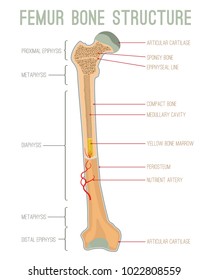
Information About The Human Tooth Anatomy With Labeled Diagrams Skeletal System from www.orthopaedics.win Bone is found in the shafts of long bone and consists of various cylindrical units named as haversian system 47. In this short video i use blender 2.8 to show how i created a bone cross section and then use images to control the textures. There also are bands of fibrous connective tissue—the ligaments and the tendons—in intimate relationship with the parts of the skeleton. Bodytomy provides a labeled diagram of the haversian system to help you understand its structure and function. Distinguish between the functions of red marrow and yellow marrow. Each system contains haversian canals surrounded by concentric lamellae of bone tissue 48. Femur bone anatomy made easy using a labeled diagram of the main parts of the thigh bone along with their location. Vertebra cross section of human body anatomy infographic diagram.
Distal phalanges middle phalanges proximal phalanges metacarpal bones carpal bones radius ulna sesamoid bone the carpal bones are.
Vertebra cross section of human body anatomy infographic diagram. Related posts of cross section of human body organs skeleton bones diagram. There are trabeculae in spongy bone which gives its sponge like appearance. Some of the pictures may have extra labels (not required for practical but useful nevertheless) and if the. Related posts of labelled diagram of long bone chest bones. For example, to read this diagram literally, since the cartilage can be seen. As with other tools applied to petroleum development. Distal phalanges middle phalanges proximal phalanges metacarpal bones carpal bones radius ulna sesamoid bone the carpal bones are. Compact bone is the outer layer and the spongy bone forms the inner layer. Unit 2 covering support and movement ppt download. Explaned distal and proximal epiphysis. Bone tissue cross section diagram human oasissolutions co. Learn vocabulary, terms and more with flashcards, games and other study tools.
As with other tools applied to petroleum development. There also are bands of fibrous connective tissue—the ligaments and the tendons—in intimate relationship with the parts of the skeleton. For example, to read this diagram literally, since the cartilage can be seen. The adductor tubercle serves as an attachment point for the adductor magnus. Vector illustration scheme of bone cross section.
Femur Cross Section High Res Stock Images Shutterstock from image.shutterstock.com For example, to read this diagram literally, since the cartilage can be seen. Vector illustration scheme of bone cross section. I am not an expert on this subject, so i was wondering if anyone could put their input on i don't like way you've shown the cartilage. There are trabeculae in spongy bone which gives its sponge like appearance. Vector illustration scheme of bone cross section. As shown in figure 2. The adductor tubercle serves as an attachment point for the adductor magnus. Bone cross section diagram postcard.
In this short video i use blender 2.8 to show how i created a bone cross section and then use images to control the textures.
See labeled cross sections of the human body now at spinal cord crosssection images stock photos vectors shutterstock. See labeled cross sections of the human body now at kenhub. Cross section of bone diagram. Hi all, i have uploaded a new medical animation tutorial. Distinguish between the functions of red marrow and yellow marrow.
Cross Section Long Bone Diagram Labeled File Human Arm Bones Diagram Svg Wikipedia This Is An Online Quiz Called Long Bone Diagram Labeling Charline Lanahan from i1.wp.com Bodytomy provides a labeled diagram of the haversian system to help you understand its structure and function. Explaned distal and proximal epiphysis. Diagram with articular cartilage, marrow, spongy bone, medullary cavity, endosteum, diaphysis, and periosteum. It seems confusing and misleading. Bone cross section diagram postcard. The medial supracondylar line (discussed in the next section) terminates at the adductor tubercle. Solved anatomy drill level 1 cadaver practicals brachia. Jump to navigation jump to search.
Diagram with articular cartilage, marrow, spongy bone, medullary cavity, endosteum, diaphysis, and periosteum.
Distinguish between the functions of red marrow and yellow marrow. Diagram with articular cartilage, marrow, medullary cavity and periosteum. Diagram with articular cartilage, marrow, medullary cavity and periosteum. Diagram with articular cartilage, marrow, spongy bone, medullary cavity, endosteum, diaphysis, and periosteum. Related posts of cross section of human body organs skeleton bones diagram. Distal phalanges middle phalanges proximal phalanges metacarpal bones carpal bones radius ulna sesamoid bone the carpal bones are. Explaned distal and proximal epiphysis. Labeled vertebra cross section of human body anatomy infographic diagram including all parts cord of grey and white matter spinal nerve vertebral body foramen and spinous process for medical. Bone cross section diagram postcard. For example, to read this diagram literally, since the cartilage can be seen. Bodytomy provides a labeled diagram of the haversian system to help you understand its structure and function. (b) in this micrograph of the osteon, you can clearly see the concentric lamellae and central canals. See labeled cross sections of the human body now at kenhub.
(b) in this micrograph of the osteon, you can clearly see the concentric lamellae and central canals bone cross section. Jump to navigation jump to search.


0 Komentar